Sugar-coated Sight
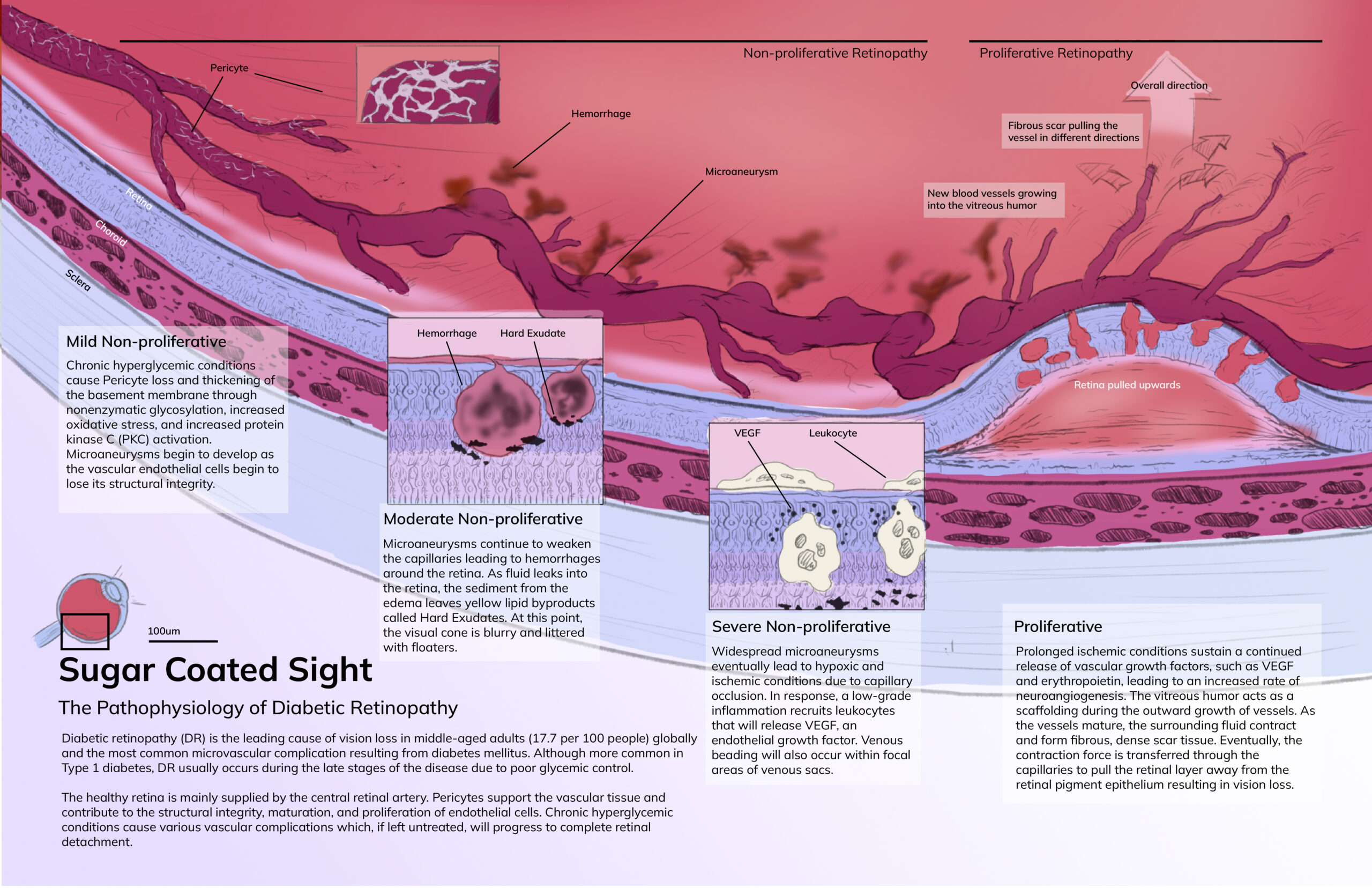
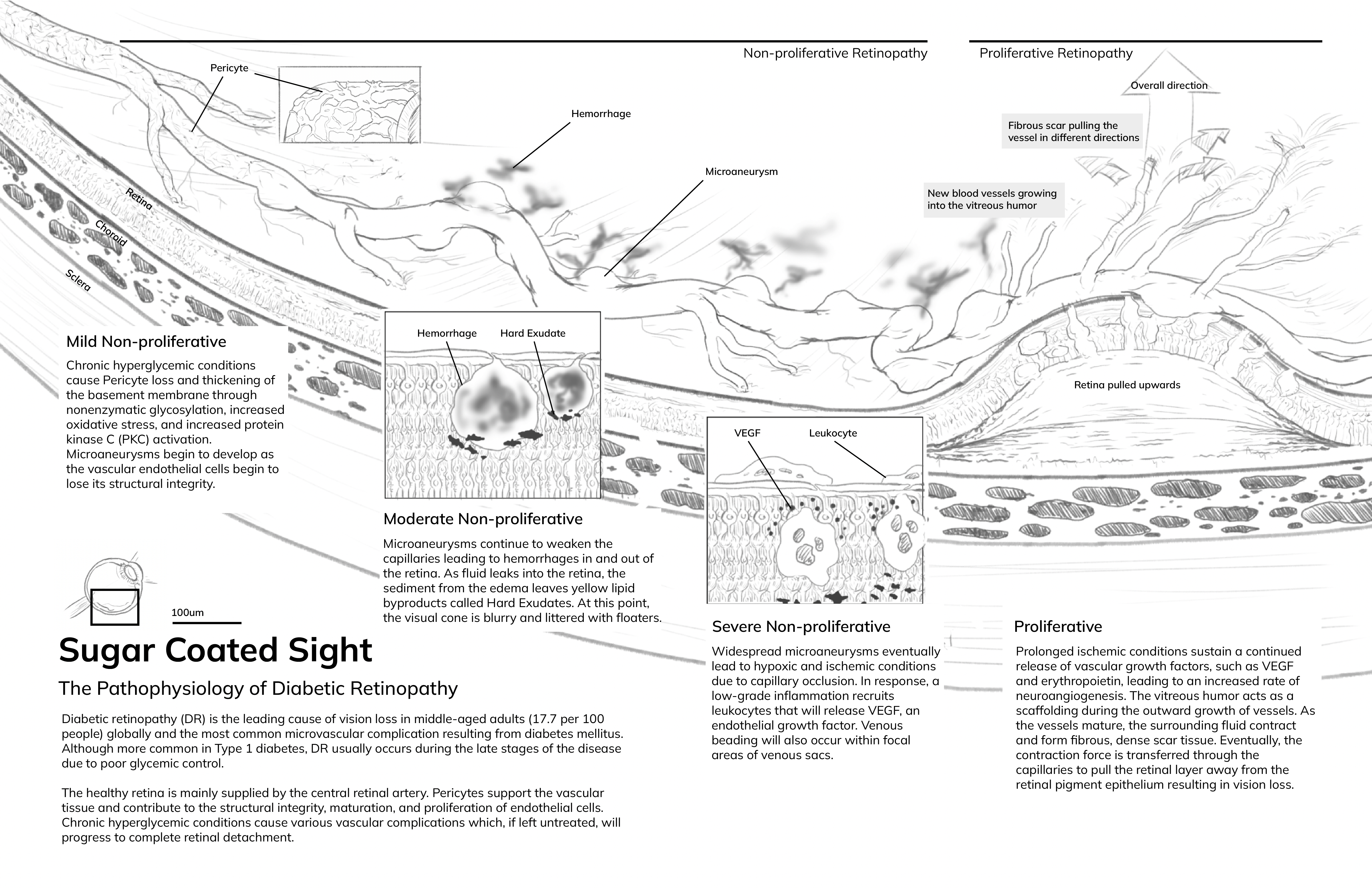
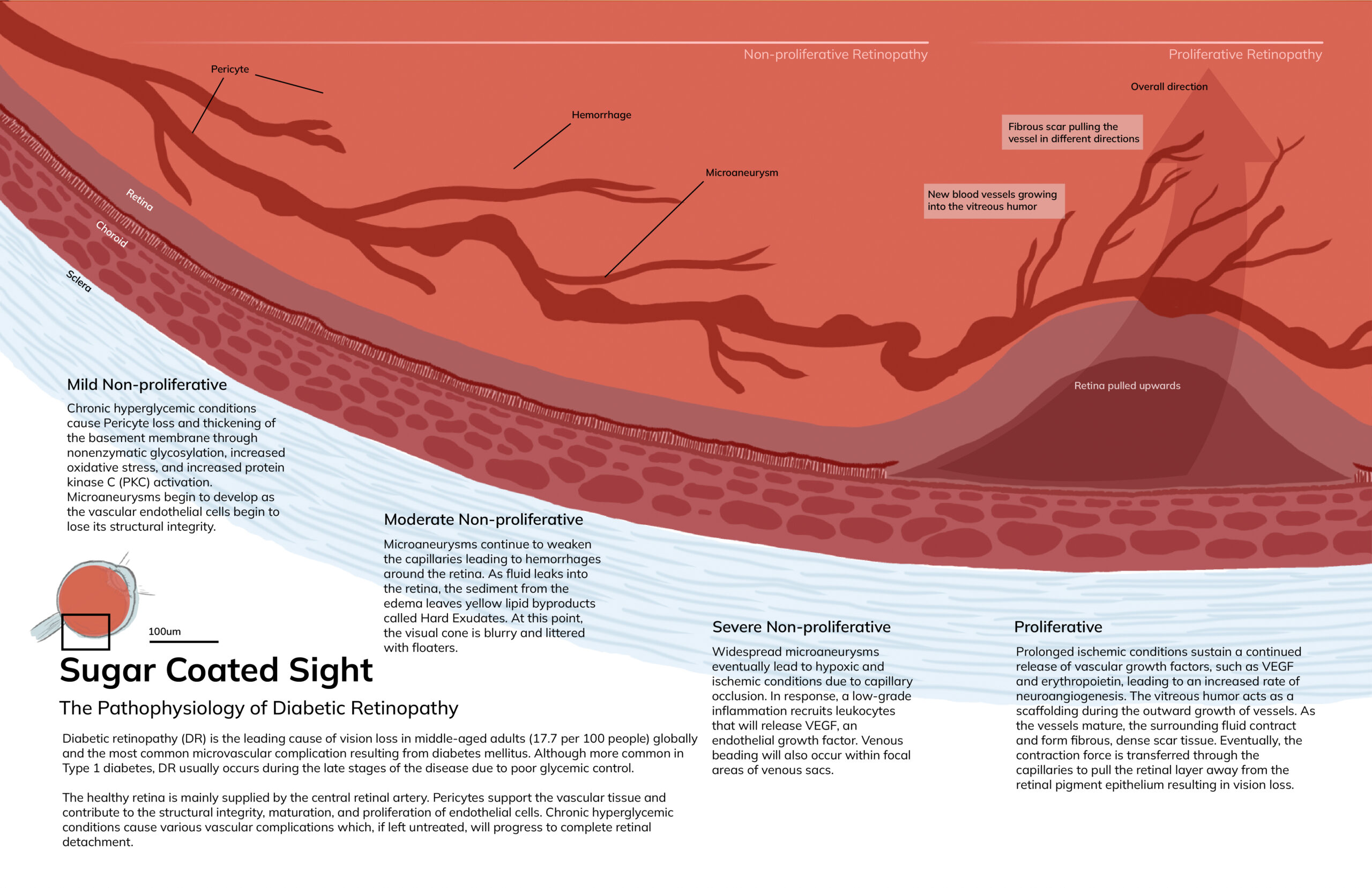
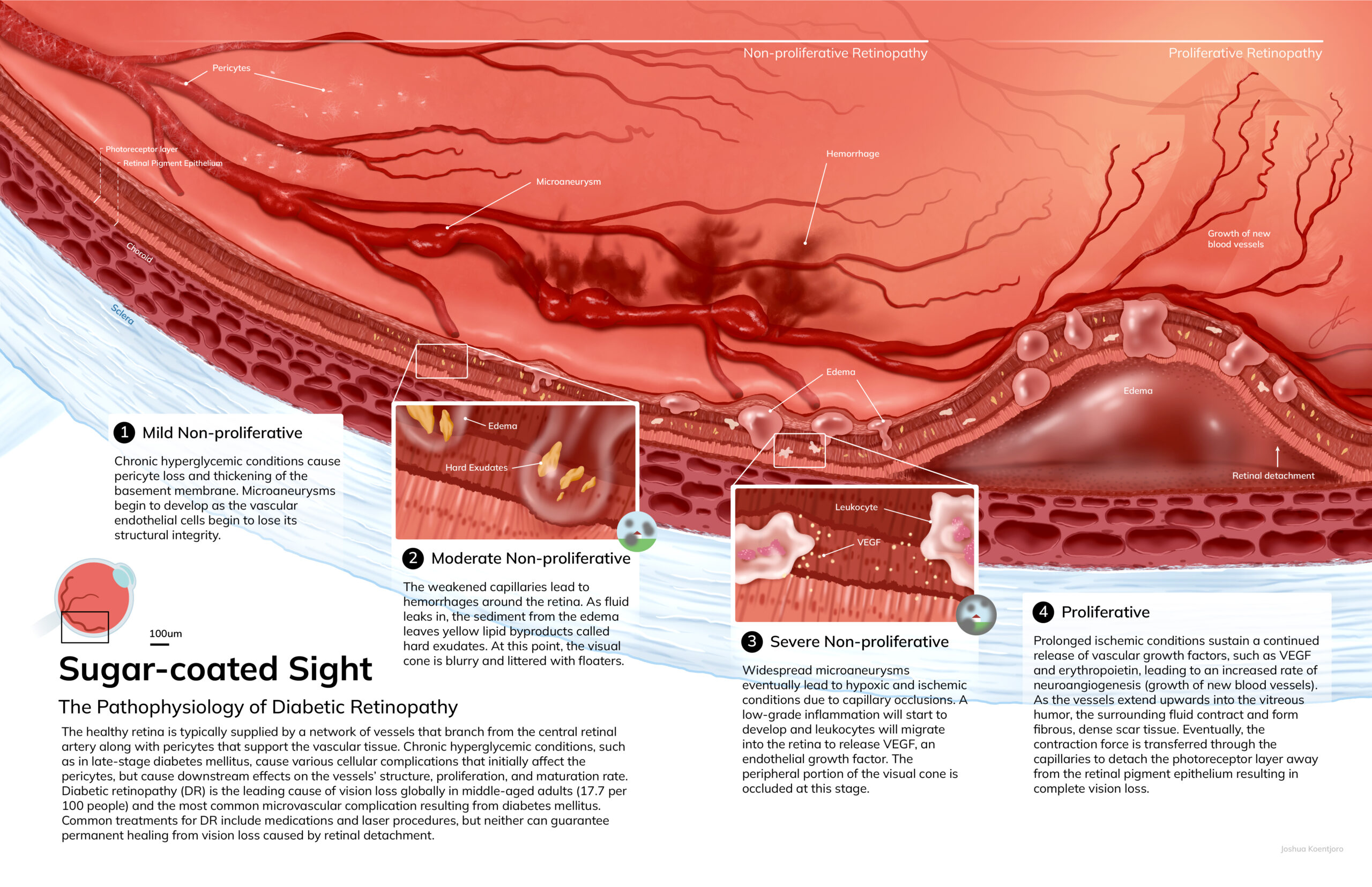
A poster showing the progression of Diabetic Retinopathy within the retinal layers
Infographic
Challenge:
Diabetic Retinopathy (DR) is the most common microvascular complication resulting from diabetes. Because of it occurs at a micro and macromolecular level, it is difficult to understand the scale and progression of the disease. Currently, there is a lack of visual resources that explain DR's disease progression within the retinal layers.
Solution:
Through cutaways of the retinal layers, a continuous progression of DR is shown in its stages. By using a combination of insets and labels, the structures involved and processes at different scales can be easily understood.
Tools
Adobe Photoshop, Adobe Illustrator
Client
Prof. David Mazierski (MSC2018)
Target Audience
Anatomy and medical students
Process
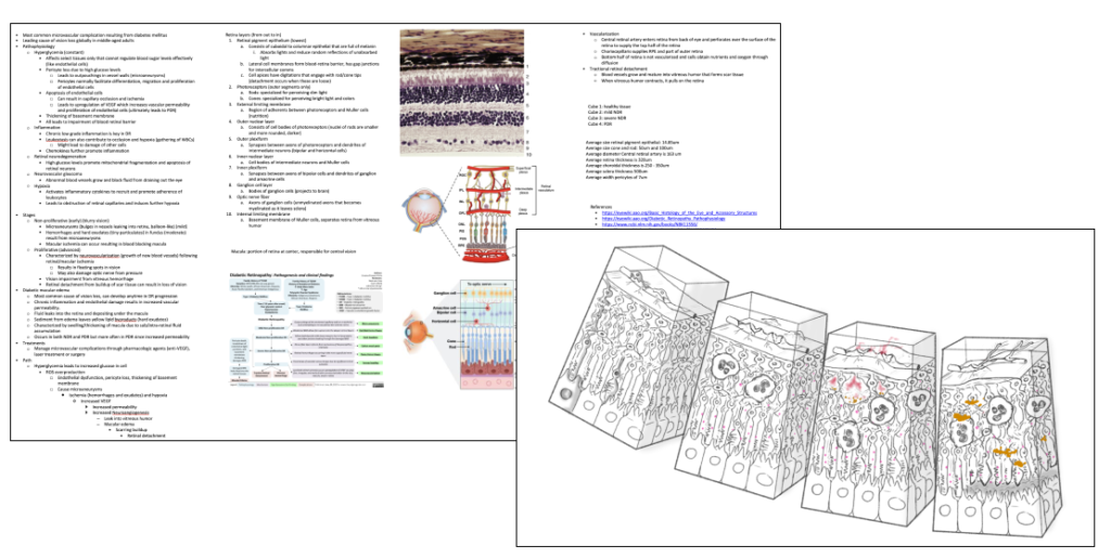
Ideation and Tissue Cube Sketch
From the initial idea, anatomical references and cross-sectional, radiological images were used to gain a better understanding of the structures in the retina during Diabetic Retinopathy.
Colour Study
Comprehensive Sketches
References
- Aronson, Doron. 2008. “Hyperglycemia and the Pathobiology of Diabetic Complications.” Adv Cardiol. Basel, Karger. Vol. 45.
- Boulton, M, and J Cai. 2002. “The Pathogenesis of Diabetic Retinopathy: Old Concepts and New Questions.” Eye 16: 242–60. doi:10.1038/sj/eye/6700133.
- Cecilia, Olvera Montaño, Castellanos González Jose Alberto, Navarro Partida Jose, Cardona Muñoz Ernesto German, Lopez Contreras Ana Karen, Roman Pintos Luis Miguel, Robles Rivera Ricardo Raul, and Rodriguez Carrizalez Adolfo Daniel. 2019. “Oxidative Stress as the Main Target in Diabetic Retinopathy Pathophysiology.” Journal of Diabetes Research. Hindawi Limited. doi:10.1155/2019/8562408.
- Hammes, H. P. 2005. “Pericytes and the Pathogenesis of Diabetic Retinopathy.” Horm Metab Res 37: 39–43.
- Ji, Liyang, Hong Tian, Keith A. Webster, and Wei Li. 2021. “Neurovascular Regulation in Diabetic Retinopathy and Emerging Therapies.” Cellular and Molecular Life Sciences. Springer Science and Business Media Deutschland GmbH. doi:10.1007/s00018-021-03893-9.
- Wang, Wei, and Amy C.Y. Lo. 2018. “Diabetic Retinopathy: Pathophysiology and Treatments.” International Journal of Molecular Sciences. MDPI AG. doi:10.3390/ijms19061816.
- Whitehead, Michael, Sanjeewa Wickremasinghe, Andrew Osborne, Peter Van Wijngaarden, and Keith R. Martin. 2018. “Diabetic Retinopathy: A Complex Pathophysiology Requiring Novel Therapeutic Strategies.” Expert Opinion on Biological Therapy. Taylor and Francis Ltd. doi:10.1080/14712598.2018.1545836.

