
Muscles and Neurovasculature of the Cubital Fossa
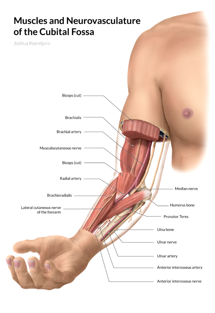
An anatomical reference using cutaways to reveal the hidden structures in the elbow
Anatomical
Challenge:
The cubital fossa has a myriad of neurovasculature structures running in different directions to different targets. In conventional textbook illustrations, they are rarely depicted relative to landmark musculature making it difficult to understand their course.
Solution:
By using strategic cutaways of the muscle layers, nerves and vessels running below are revealed in their course towards their target.
Tools
Cinema 4D, Adobe Photoshop, Adobe Illustrator
Client
Prof. Jodie Jenkinson (MSC2023H)
Content Advisor
Prof. Carol Heck (University of Toronto)
Target Audience
Anatomy and medical students
Muscles and Neurovasculature of the Cubital Fossa
An anatomical reference using cutaways to reveal the hidden structures in the elbow
Anatomical
Challenge:
The cubital fossa has a myriad of neurovasculature structures running in different directions to different targets. In conventional textbook illustrations, they are rarely depicted relative to landmark musculature making it difficult to understand their course.
Solution:
By using strategic cutaways of the muscle layers, nerves and vessels running below are revealed in their course towards their target.
Tools
Cinema 4D, Adobe Photoshop, Adobe Illustrator
Client
Prof. Michael Corrin (MSC2001Y)
Content Advisor
Prof. Carol Heck (University of Toronto)
Target Audience
Anatomy and medical students
Process
Ideation and Research
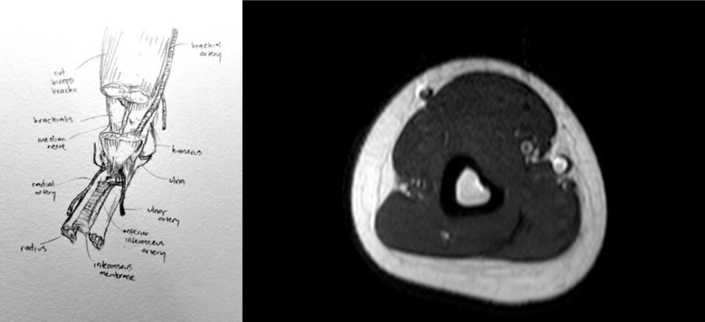
From the initial idea, anatomical references and cross-sectional, radiological images were used to gain a better understanding of the structures in the cubital fossa area.
Maquettes
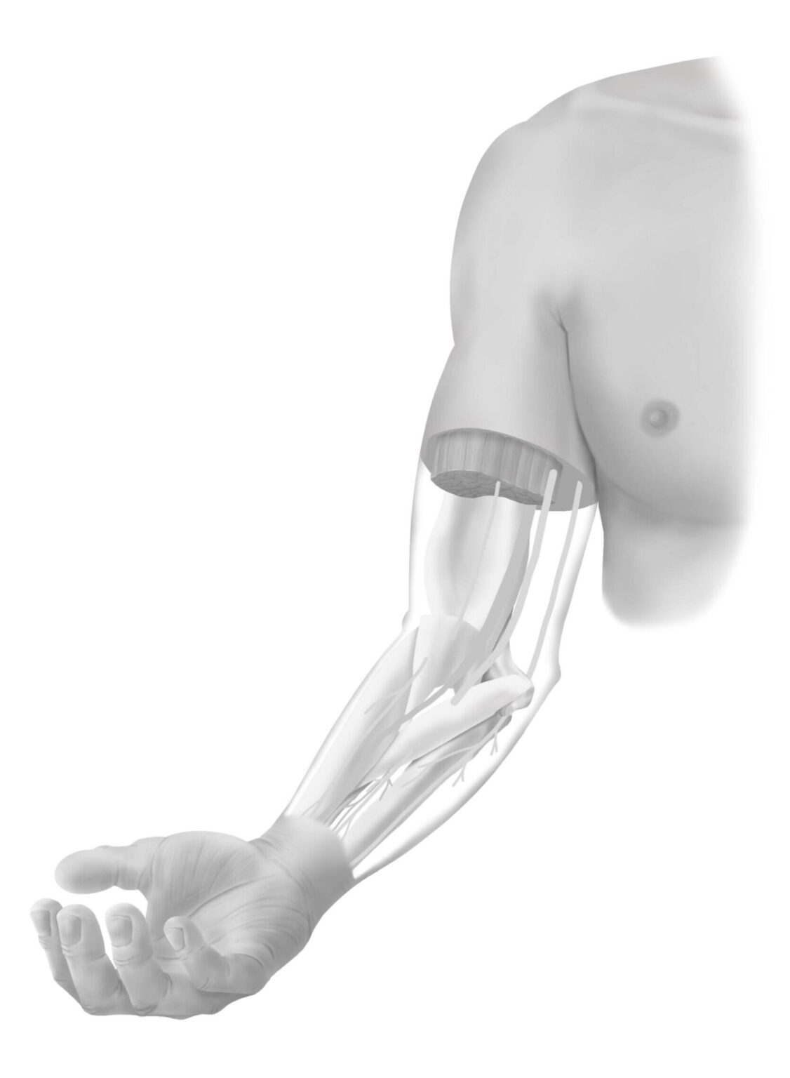
Using OBJ files of the arm bones as the base, Boolean functions were utilized to sculpt the arm, major muscles, and main neurovasculature structures within Cinema 4D.
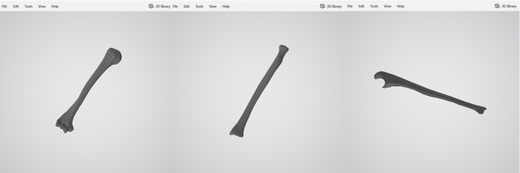
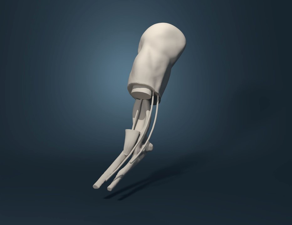
Maquettes
Using OBJ files of the arm bones as the base, Boolean functions were utilized to sculpt the arm, major muscles, and main neurovasculature structures within Cinema 4D.


Comprehensive Sketches
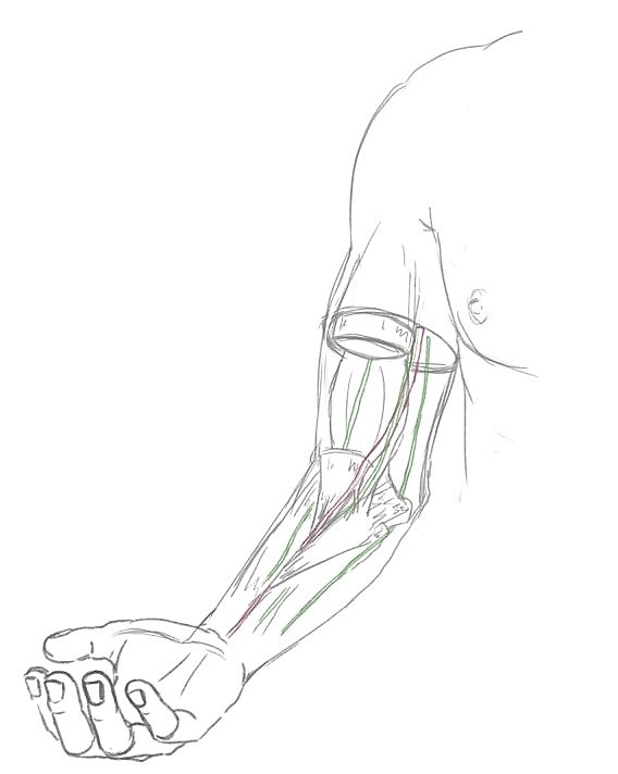
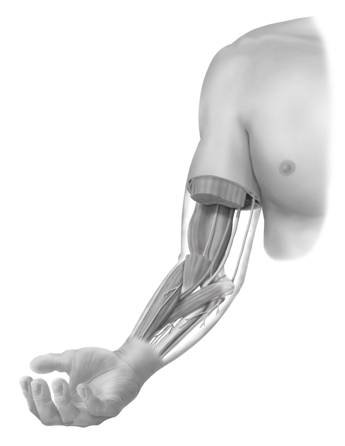
Using the maquettes, comprehensive sketches of the arm are created. The hand was added to add additional context to the illustration.


Render and Compiling
By starting with a grayscale copy in Adobe Photoshop, contrast and form was the main focus before colorizing the illustration. Afterwards, labels were added in Adobe Illustrator.

References
- Annals of Cardiothoracic Surgery (ACS) (2015) Open thoracoabdominal aortic aneurysm repair. [Video]. YouTube. https://www.youtube.com/watch?v=ngr80zG8-WI (Accessed December 20, 2021)
- Agur, A. M. R., Dalley, A. F. (2017). Grant’s Atlas of Anatomy (14th ed, pp. 70, 76, 111, 129-130, 142-144). Wolters Kluwer.
- Bo, W. J. (2007). Basic atlas of sectional anatomy: With correlated imaging. (2nd ed, pp. 340, 342, 347) Saunders Elsevier.
- Cleveland Clinic. (2021). Coronary Artery Bypass Surgery: Internal Mammary Arteries (Graphic) [Video]. YouTube. https://www.youtube.com/watch?v=PO6ioZXVBvs (Accessed December 20, 2021)
- Majid, A., Majid D. (2016). M&M Essential Anatomy (3rd ed, pp. 55, 174). Pearson Learning Solutions.
- Micheau, A., Hoa, D. (2021). MRI of the upper extremity anatomy – Atlas of the human body using cross-sectional imaging. https://www.imaios.com/en/e-Anatomy/Upper-Limb/Upper-extremity-MRI (Accessed December 20, 2021)
- Netter, F. H. (2019). Netter Atlas of Human Anatomy (7th ed, pp. 463-467, 421, 423, 426-430, 436, BP100-101). Saunders Elsevier.
- Standring, S., Gray, H. (2008). Gray’s Anatomy: The Anatomical Basis of Clinical Practice (40th ed, pp. 838-840, 845) Saunders Elsevier.
- Stryker. (2015). Stryker Trauma & Extremities | VariAx Elbow Locking Plate System | 180 Degree Plating [Video]. YouTube. https://www.youtube.com/watch?v=sUIVjcTgW90&t=430s (Accessed December 20, 2021)
- Tatco, V. Normal MRI of the arm. https://radiopaedia.org/cases/normal-mri-of-the-arm (Accessed December 20, 2021)